FAQ
Register
Login

The Journal Of Heart Valve Disease
Open Access
Menu
Submit your article
The Journal of Heart Valve Disease
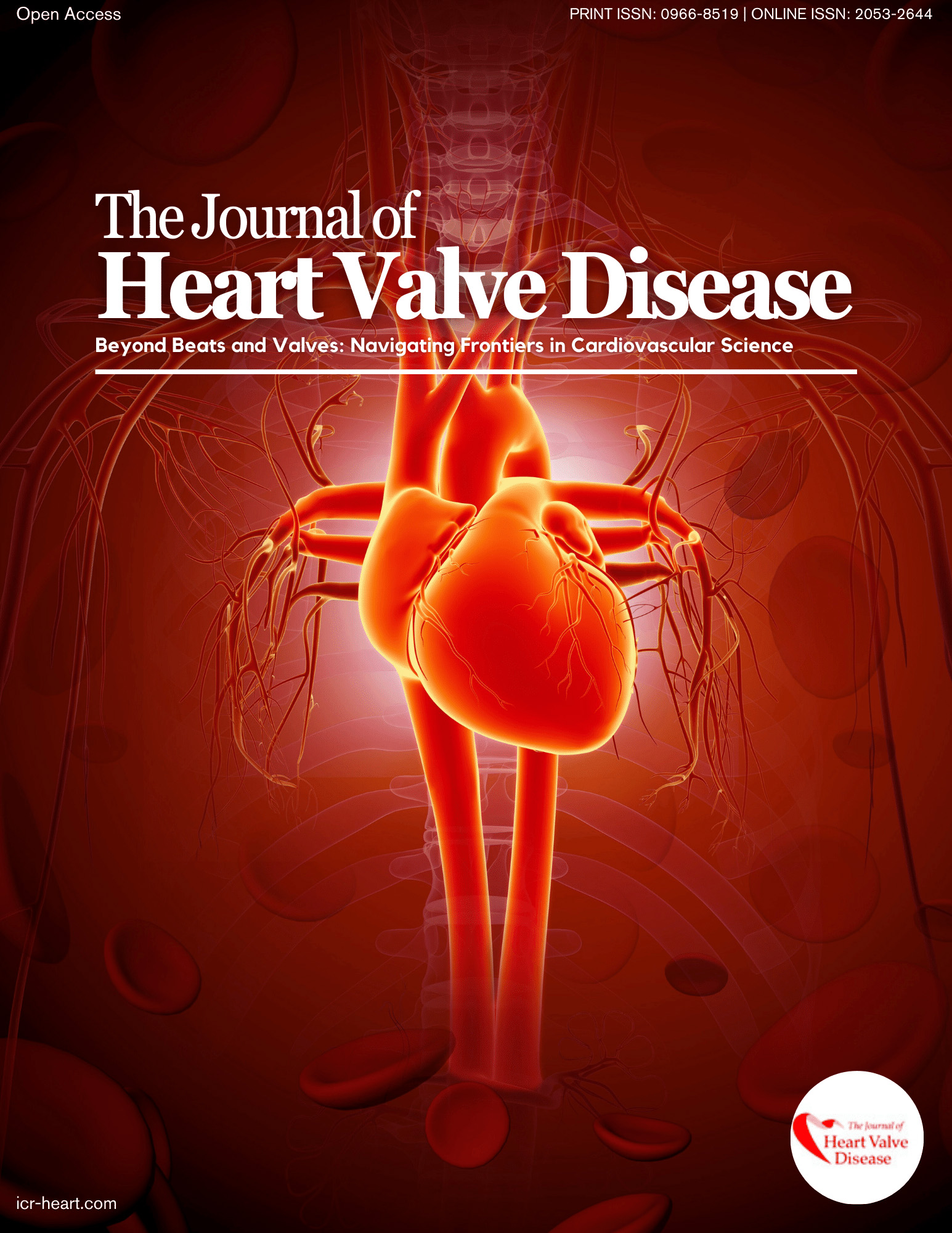
Established in 1992, The Journal of Heart Valve Disease has positioned itself as a key player in the heart valve disease sector. Our core mission is to facilitate global collaboration across various disciplines, transcending geographical boundaries to advance the understanding and treatment of this prevalent medical condition. In a remarkable feat, we earned a coveted spot on Index Medicus within just 18 months of our inception, solidifying our commitment to showcasing cutting-edge research and clinical insights in the field. Since then, we've consistently been at the forefront of disseminating valuable knowledge. Today, our reach extends to 74 countries across five continents, making us a globally recognized source of information. Over the past decade, we've proudly published contributions from clinicians, academics, research scientists, industry experts, and regulatory bodies in 52 countries spanning five continents. We actively seek and welcome a wide range of submissions, including original research publications, thought-provoking editorials, comprehensive reviews, technical papers covering surgery, cardiology, and fundamental science, illustrative case reports, captivating "Image of the Month" features, engaging correspondence, and informative book reviews. Print ISSN: 0966-8519 Online ISSN: 2053-2644

